EDIT Lab / LSU

Three research directions.

Molecules binding at an engineered electrode. Whole blood changing as it clots. Both of those measured while the sample is moving.

Research

What we work on.

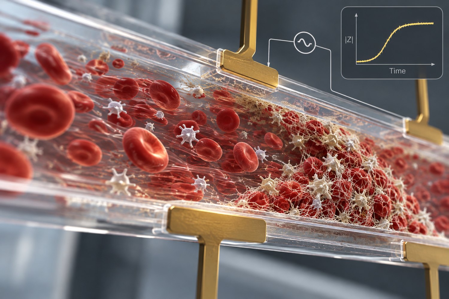
Microfluidic channel with electrodes above and below; red blood cells and platelets flow left to right while a fibrin-rich thrombus builds downstream.
Blood under flow in an electrode-instrumented channel, with thrombus accumulating downstream. Illustration.

Coagulation under flow

Blood does not clot the same way at rest as it does under shear, yet most routine clinical coagulation assays are performed under static or near-static conditions. We build microfluidic sensors that hold blood under controlled flow and track its electrical response, so shear becomes something we set and vary.

The clinical problem is extracorporeal support. Patients on ECMO or continuous renal replacement therapy sit between bleeding and circuit thrombosis, and the measurements clinicians have give an intermittent and incomplete picture of the hemostatic state driving both. We are working toward a sensor that sits closer to the circuit and runs continuously.

  • How does shear rate reshape the electrical signature of clot formation?
  • Can emerging thrombotic changes be detected before they produce measurable circuit dysfunction?
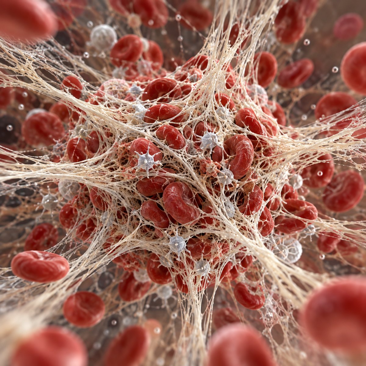
Dense fibrin fibers forming a three-dimensional mesh around red blood cells and activated platelets.
A fibrin network enmeshing red cells and platelets, the structure our measurements track as it forms and contracts. Illustration.

Dielectric sensing of whole blood

Between kilohertz and megahertz, the electrical properties of whole blood change as cells and fibrin reorganize into a clot. We use that to study clot contraction, the platelet-driven phase that clot-time assays miss.

We also test how sensor geometry, chamber height and electrode configuration in particular, affects sensitivity to clot formation and contraction. When a sensor shows no signal, we want to know whether the biology was quiet or the chamber was wrong.

  • Which electrical features map onto contractile behavior, and over what range?
  • How do inflammatory and uremic environments alter contraction independently of clot time?
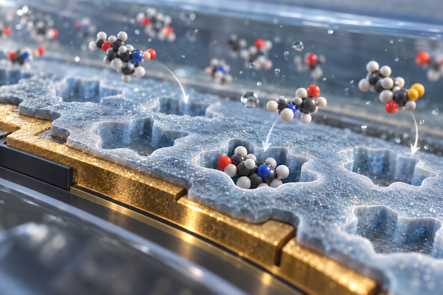
Gold electrode coated with an imprinted polymer film; shaped cavities sit in the film, one occupied by a matching molecule while differently shaped molecules remain in solution.
An imprinted polymer on a gold electrode: cavities shaped to the target, with non-matching molecules passing by. Illustration.

Electrochemical biosensing

We engineer nanomaterial-modified and molecularly imprinted electrode interfaces to detect circulating biomarkers associated with kidney and cardiovascular disease. Current work focuses on protein-bound uremic toxins, which accumulate in chronic kidney disease, are incompletely cleared by conventional dialysis, and may contribute to cardiovascular complications.

The same interface-engineering work extends to other samples: chemiresistive gas sensing for ammonia, and sorting adulterated milk from its impedance spectrum. The transduction differs in each case. What carries over is designing the sensing surface around the specific target.

  • How can electrode chemistry provide selective detection of uremic toxins in complex biological samples?
  • Can one cartridge report a molecular target and its functional consequence?
Capabilities

What the lab can measure.

Broadband bioelectrical measurement

We measure impedance from kilohertz to megahertz, across multiple channels in parallel, and repeatedly over time. A clotting experiment gives us a curve, not a single endpoint.

Microfluidic models of hemostasis

Flow rate, shear, surface coating, and coagulation activation can each be set and varied independently. That matters because it lets us separate what the sensor is doing from what the blood is doing.

Sensor and interface engineering

The sensors we build, capacitive microfluidic chips, PCB electrode structures, and 2D material and molecularly imprinted electrodes, are designed around the measurement rather than adapted from something general.

Signal analysis

We pull features tied to clot initiation, fibrin formation, and contraction, and check them against conventional coagulation assays. When the relevant signal is distributed across frequency and time, we use machine learning to locate it.

Platform evolution

ClotChip was introduced during Dr. Maji's doctoral research at Case Western Reserve University, as a microfluidic dielectric sensing platform for whole blood coagulation. That work established that an electrical measurement can capture clinically meaningful changes in hemostasis from a drop-scale sample.

At LSU we are taking that foundation in our own directions: coagulation under controlled flow, blood-contacting extracorporeal circuits, simpler sensor architectures that are easier to manufacture, and electrochemical detection of circulating biomarkers in cardiovascular and kidney disease.

Foundational platform: Maji et al., IEEE Transactions on Biomedical Circuits and Systems, 11(6), 1459–1469, 2017.